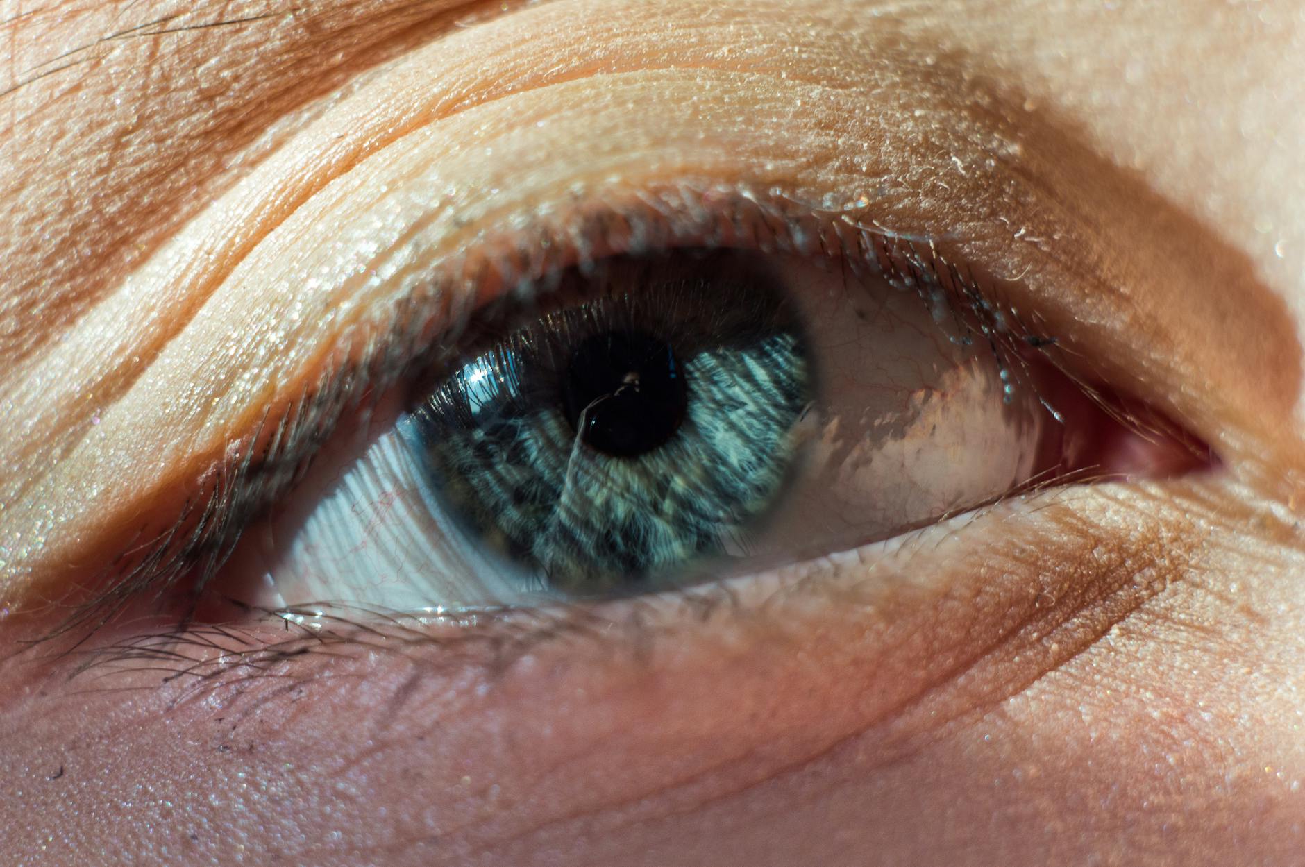
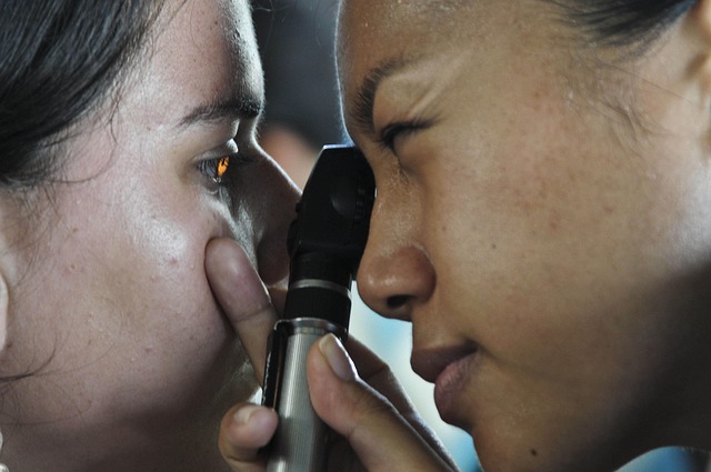
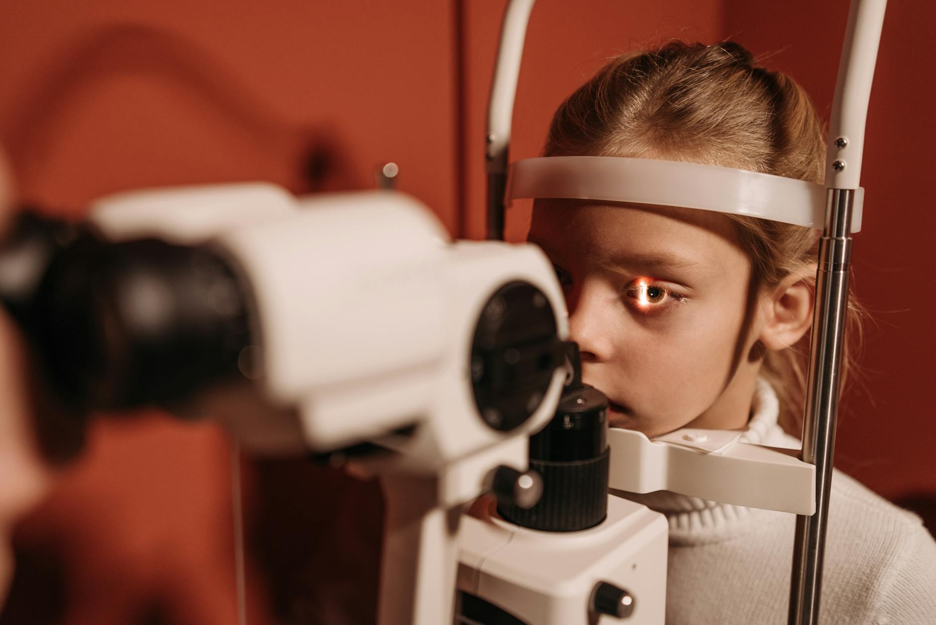
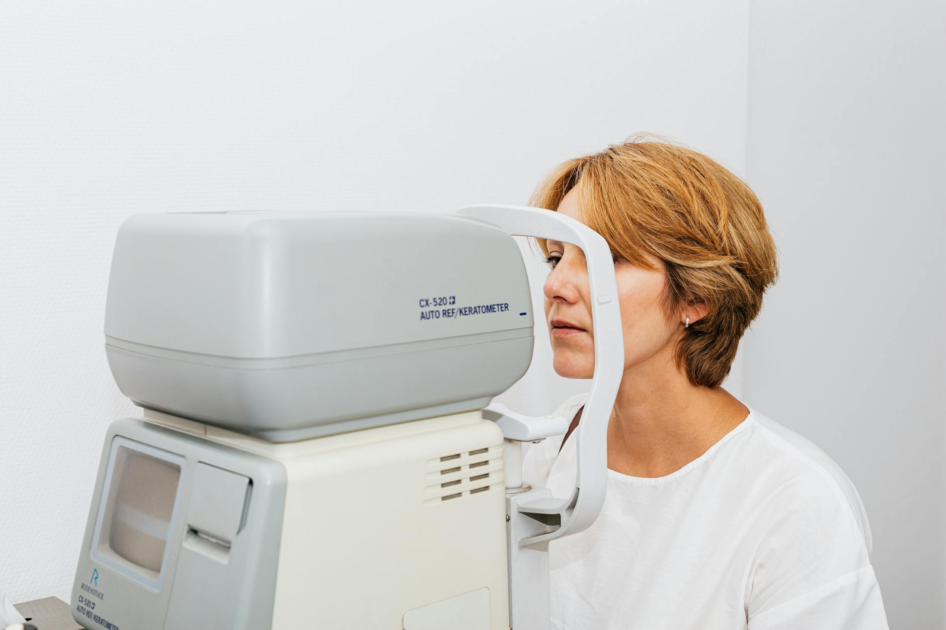
Maintaining optimal vision health requires vigilance regarding subtle and obvious changes in your daily sight. Many serious ocular conditions present early warning signs that are frequently ignored until significant damage has occurred. Regular examinations serve as the first line of defense against preventable vision loss and systemic health issues often detectable through the eyes. Paying close attention to these forty indicators can ensure timely medical intervention and preserve your quality of life. The following guide outlines key symptoms and situations that warrant a prompt professional consultation.
Sudden Blurry Vision

A rapid decrease in visual clarity often indicates a significant change in your eye health that requires urgent attention. This symptom might affect just one eye or both simultaneously depending on the underlying cause. It can stem from issues ranging from a simple prescription change to more serious conditions like a detached retina or stroke. Ignoring this sudden shift puts you at risk for permanent vision loss or unaddressed systemic health problems. Immediate professional evaluation helps determine the root cause and appropriate treatment plan.
Persistent Double Vision
Seeing two images of a single object creates confusion and indicates a misalignment or neurological issue affecting the eyes. This condition can occur in one eye alone or only when both eyes are open. Causes may include corneal irregularities or problems with the muscles controlling eye movement. It can also signal serious neurological events like a brain aneurysm or stroke. A comprehensive exam is necessary to rule out life-threatening conditions and restore single binocular vision.
New Floaters
Small specks or cobwebs drifting across your field of vision are common but a sudden increase warrants concern. These shapes are actually shadows cast on the retina by clumps of gel within the vitreous humor. While often benign they can sometimes signal a retinal tear or detachment requiring emergency surgery. Noticing a shower of these spots suggests the vitreous may be pulling away from the back of the eye. Prompt assessment prevents potential blindness associated with untreated retinal damage.
Flashes of Light

Brief streaks or lightning bolts of light in your peripheral vision suggest traction on the retina. These photopsias often occur when the vitreous gel rubs or pulls against the light-sensitive retinal tissue. This symptom frequently accompanies new floaters and serves as a warning sign for retinal detachment. It can also be associated with ocular migraines even without a headache present. Differentiating between a migraine and a retinal emergency requires a dilated eye examination.
Severe Eye Pain

Sharp or throbbing pain in the eye is never normal and suggests an acute injury or infection. This discomfort might stem from a scratched cornea or a foreign object lodged beneath the eyelid. Intense pain accompanied by nausea could indicate narrow-angle glaucoma which constitutes a medical emergency. Ignoring this symptom leads to unnecessary suffering and potential permanent damage to ocular structures. Seeing a specialist ensures the pain is managed and the underlying injury is treated.
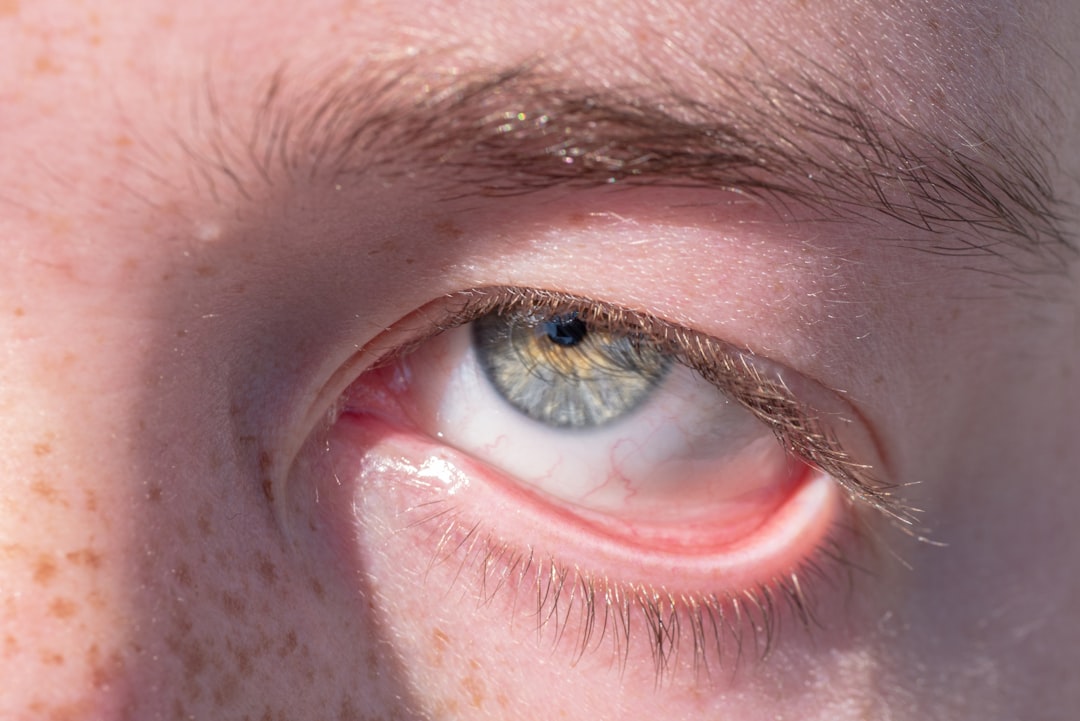
Persistent Redness
Eyes that remain red or bloodshot for an extended period indicate inflammation or infection needing treatment. This discoloration occurs when small blood vessels on the surface of the eye expand due to irritation. It acts as a hallmark sign for conditions like conjunctivitis or uveitis. Over-the-counter drops often mask the symptom without curing the root infection or allergy. A doctor can prescribe specific medications to clear the redness and resolve the underlying issue.
Chronic Dry Eyes

A constant gritty or sandy sensation suggests your eyes are not producing enough quality tears for lubrication. This condition causes significant discomfort and fluctuates with environmental factors like wind or air conditioning. Long-term dryness damages the corneal surface and increases susceptibility to infections. Artificial tears provide temporary relief but fail to address the dysfunction of the tear glands. Professional treatments can unblock oil glands and restore a healthy tear film.
Excessive Tearing
Watering eyes often paradoxically signal dryness as the eye overcompensates with reflex tears. This overflow can also result from blocked tear ducts that prevent proper drainage. Constant tearing blurs vision and causes skin irritation around the eyelids. It may also indicate an allergic reaction or a response to a foreign body. An eye doctor can flush the ducts or prescribe medication to balance tear production.
Extreme Light Sensitivity

Finding normal indoor lighting or sunlight painful indicates a condition known as photophobia. This symptom frequently accompanies migraines or severe dry eye syndrome. It can also signal inflammation inside the eye such as iritis or uveitis. Squinting constantly in regular light leads to headaches and eye strain. Evaluating this sensitivity helps diagnose infections or corneal abrasions that require protection.
Night Blindness

Difficulty seeing in low-light conditions or while driving at night suggests a problem with the rod cells in the retina. This issue makes transitioning from bright to dark environments dangerously slow. It serves as an early indicator for cataracts or retinitis pigmentosa. Vitamin A deficiency is a rare but possible cause for this reduction in night vision. Addressing this symptom is crucial for safety during evening activities and travel.
Rainbow Halos Around Lights

Seeing colored rings around streetlights or headlights often points to swelling in the cornea. This visual phenomenon is a classic symptom of acute angle-closure glaucoma. The fluid pressure builds up rapidly and clouds the front surface of the eye. It requires immediate medical intervention to lower intraocular pressure and prevent optic nerve damage. Ignoring these halos can lead to rapid and irreversible vision loss.
Frequent Headaches

Recurring pain in the head or brow area often stems from uncorrected refractive errors. Your eyes strain to focus which creates tension in the surrounding muscles. This symptom typically worsens after visual tasks like reading or computer use. Updating your eyeglass or contact lens prescription relieves this unnecessary burden on the visual system. A comprehensive exam rules out ocular causes for chronic head pain.
Constant Squinting

Narrowing the eyes to see clearly is a subconscious attempt to improve focus by changing the eye shape. This habit indicates a refractive error such as myopia or astigmatism that needs correction. It leads to premature wrinkles and significant muscle fatigue around the face. Children often display this behavior when they cannot see the board at school. Corrective lenses easily resolve the need to squint and relax the facial muscles.
Digital Eye Strain

Prolonged screen time leads to symptoms like fatigue and blurring known as computer vision syndrome. The eyes blink less frequently when staring at digital devices causing surface dryness. This condition reduces productivity and causes discomfort during the workday. Special computer glasses or vision therapy can alleviate the stress placed on the focusing system. An optometrist can recommend specific ergonomic adjustments and lens coatings.
Eyelid Twitching

Involuntary spasms of the eyelid muscle are annoying and often linked to stress or fatigue. While usually harmless persistent twitching can signal neurological issues or severe dry eye. Excessive caffeine intake or lack of sleep exacerbates these muscle contractions. If the twitching spreads to other parts of the face it requires neurological evaluation. Addressing lifestyle factors usually resolves minor cases of this distraction.
Droopy Eyelids
Ptosis or the drooping of the upper eyelid can obstruct vision and indicate muscle weakness. This condition might develop slowly with age or appear suddenly due to nerve damage. It can sometimes signal a serious underlying condition like myasthenia gravis or a brain tumor. Surgical intervention is often necessary to lift the lid and clear the visual field. An examination determines if the cause is muscular or neurological in nature.
Bulging Eyes

Eyes that appear to protrude from the sockets suggest thyroid eye disease or Graves’ disease. This physical change occurs due to swelling of the fat and muscles behind the eye. It often leads to dryness as the eyelids cannot close completely during sleep. Severe cases result in optic nerve compression and vision loss. Managing the underlying thyroid condition helps stabilize the ocular appearance and health.
Yellowing of the Whites
A yellowish tint to the sclera is a primary sign of jaundice and liver dysfunction. This visible change indicates high levels of bilirubin in the bloodstream. It serves as a systemic warning rather than a purely ocular problem. Immediate medical attention is needed to assess liver function and overall health. Treating the systemic condition will typically resolve the discoloration in the eyes.
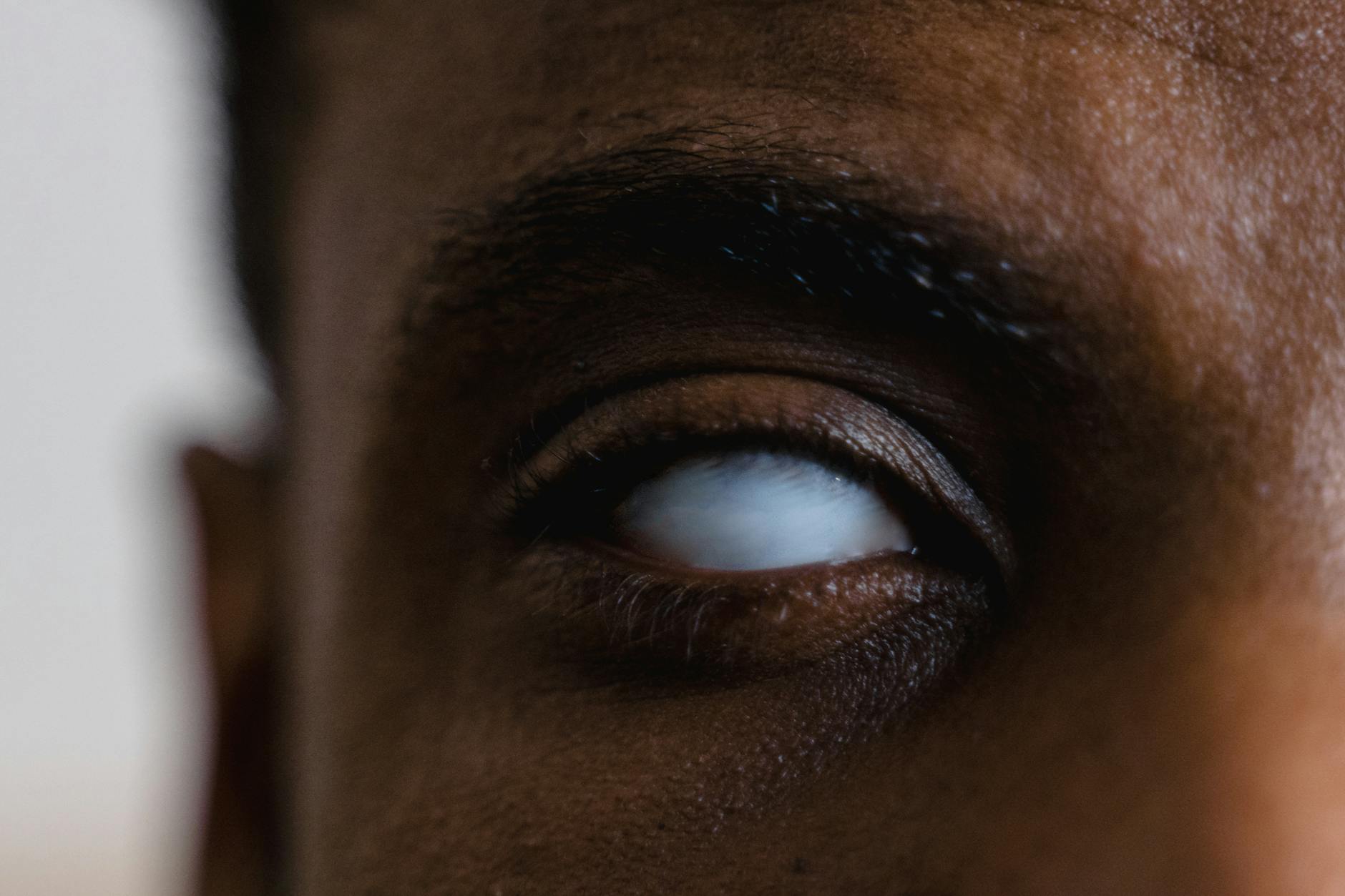
Unequal Pupil Sizes
Anisocoria or a noticeable difference in pupil size can be normal but sudden onset is alarming. It may indicate a neurological trauma or a viral infection affecting the iris. This symptom appears in cases of brain injury or aneurysm compression. Evaluation helps distinguish between benign physiological differences and life-threatening emergencies. Prompt checking of pupil reactivity is essential for diagnosis.
Loss of Peripheral Vision

Tunnel vision or the darkening of side vision is a hallmark sign of glaucoma. This sneaky disease damages the optic nerve often without pain or early warning. Losing peripheral sight increases the risk of falls and accidents during daily movement. Once lost this vision cannot be restored through any medical means. Regular pressure checks detect glaucoma early enough to halt its progression.
Distorted Vision

Straight lines appearing wavy or bent suggests fluid accumulation beneath the macula. This distortion is a key symptom of age-related macular degeneration. It significantly impacts central vision needed for reading and recognizing faces. Early detection allows for injections that can dry up the fluid and preserve sight. Use of an Amsler grid at home helps monitor for these subtle changes.
Blind Spots
A scotoma or missing area in your visual field requires immediate investigation. This dark or empty spot can indicate a retinal hole or optic nerve damage. Migraine auras often present as a temporary blind spot with shimmering edges. Persistent blind spots suggest a static issue within the eye or visual cortex. Mapping the visual field helps doctors pinpoint the location of the damage.
Difficulty Recognizing Colors

A sudden inability to distinguish colors or a fading of color intensity indicates retinal or nerve issues. This change differs from congenital color blindness which is present from birth. It can signal optic neuritis or toxic nutritional optic neuropathy. Some medications also cause color vision defects as a side effect. Testing establishes the severity and potential cause of the color desaturation.
Intense Itching

Persistent itching is the most common symptom of ocular allergies. This sensation compels rubbing which releases more histamines and worsens the condition. It can also indicate blepharitis or an infestation of eyelash mites. Anti-allergy drops provide relief more effectively than oral antihistamines. Identifying the allergen helps prevent future uncomfortable episodes.
Burning Sensation

Feeling like your eyes are burning often points to inflammation of the eyelids or surface dryness. This symptom is common in environments with smoke or chemical irritants. It can also result from the prolonged use of contact lenses. Lubricating drops and lid hygiene regimens typically soothe the burn. Chronic burning requires investigation into the quality of the tear film.
Gritty Foreign Body Sensation

Feeling like something is in your eye when nothing is visible suggests a corneal abrasion or ulcer. This sensation is highly distracting and causes excessive blinking and tearing. It often occurs after sleeping in contact lenses or getting dust in the eye. Antibiotic drops are frequently needed to prevent infection in the compromised tissue. A slit-lamp exam reveals the microscopic scratch causing the feeling.
Crusty Discharge

Waking up with eyelids stuck together indicates bacterial or viral conjunctivitis. The discharge can be yellow, green, or clear depending on the pathogen. This symptom is highly contagious and requires strict hygiene to prevent spreading. Warm compresses help loosen the crusts and provide comfort. Medical treatment shortens the duration of the infection and reduces transmission risk.
Swollen Eyelids
Puffiness or inflammation of the lids can result from allergies or fluid retention. It is also a sign of orbital cellulitis which is a serious infection spreading to the eye socket. Swelling often accompanies chalazia or sties that block the oil glands. Cold compresses reduce allergic swelling while warm ones help blocked glands. Differentiating between infection and allergy is key to proper treatment.
Recurring Sties
Frequent painful lumps on the eyelid margin suggest chronic blepharitis or poor lid hygiene. These localized infections occur at the base of the eyelashes. They can harden into cysts that require surgical removal if they do not drain. Persistent lesions should be biopsied to rule out rare eyelid cancers. A doctor can prescribe antibiotic ointments and preventative cleaning routines.
Ingrown Eyelashes
Trichiasis occurs when eyelashes grow inward and scratch the sensitive cornea. This condition causes constant irritation and increases the risk of infection. It often results from previous eye trauma or chronic inflammation. Plucking provides only temporary relief as the lashes grow back sharper. Electrolysis or cryotherapy offers a permanent solution to remove the offending follicles.
Chemical Splash

Getting household cleaners or industrial chemicals in the eye is a dire emergency. Acidic or alkaline substances can melt the cornea within minutes. Immediate flushing with water is critical before even heading to the doctor. Professional assessment determines the pH balance and depth of the burn. prompt treatment prevents extensive scarring and blindness.
Visible Object in Eye
Debris that does not wash out with tears or saline requires professional removal. Attempting to dig it out yourself can push it deeper or scratch the cornea. Metal particles can rust quickly and cause a larger ring of damage. A doctor uses a biomicroscope and sterile tools to safely extract the object. Post-removal drops ensure the wound heals without infection.
Contact Lens Intolerance

sudden discomfort while wearing lenses you previously tolerated indicates a developing issue. It often signals giant papillary conjunctivitis or a corneal ulcer. Continuing to wear the lenses exacerbates the inflammation and risks permanent scarring. You may need to switch lens materials or cleaning solutions. A break from lens wear allows the eye surface to recover fully.
Diabetic Vision Changes
Fluctuating vision in diabetics often relates to unstable blood sugar levels swelling the lens. It serves as a warning sign for diabetic retinopathy which damages retinal blood vessels. Early stages often have no symptoms making regular checks vital. Leaking vessels cause swelling and bleeding that obscures vision. Strict glucose control and laser treatments manage this leading cause of blindness.
Hypertensive Eye Signs
High blood pressure causes changes in the retinal blood vessels visible during an exam. These vascular changes often precede damage in the kidneys or heart. Arteriovenous nicking or retinal hemorrhages are specific signs observable by a doctor. Controlling systemic blood pressure prevents further ocular and organ damage. The eye exam acts as a window into your overall cardiovascular health.
Family History of Eye Disease

Having relatives with glaucoma or macular degeneration increases your genetic risk profile. Many hereditary eye diseases show no early symptoms but cause irreversible damage. Knowing your family history prompts earlier and more frequent screening schedules. Genetic testing may be appropriate for certain retinal dystrophies. Early detection remains the most effective tool for managing inherited conditions.
Trouble Focusing Near

Presbyopia naturally stiffens the lens and makes reading close up difficult after age forty. Holding menus or phones further away is the classic behavioral sign. This change is inevitable but easily corrected with reading glasses or multifocals. Ignoring it leads to headaches and eye fatigue. An updated prescription restores the ability to perform near tasks comfortably.
Difficulty Adapting to Light Changes

Struggling to see when moving from a dark room to a bright one suggests retinal stress. This delayed adaptation affects mobility and safety in variable lighting. It can indicate early macular degeneration or vitamin deficiencies. Photoreceptor cells lose their ability to regenerate pigments efficiently. Evaluation determines if dietary changes or protective eyewear can assist.
Vision Changes with Medication

Certain systemic drugs have known side effects that impact ocular health. Corticosteroids can accelerate cataract formation and increase eye pressure. Hydroxychloroquine requires monitoring for potential retinal toxicity. It is crucial to inform your eye doctor of all prescriptions you take. Regular monitoring ensures medications do not cause lasting visual harm.
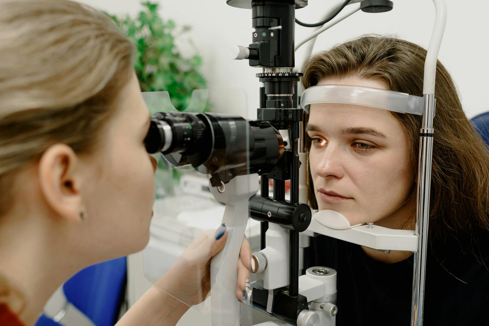
Overdue Eye Exam
Simply not having seen an eye doctor in over two years is a valid reason to go. Many conditions are asymptomatic in their early and most treatable stages. Your prescription may have drifted slightly causing low-grade strain you haven’t noticed. Routine wellness checks establish a baseline for your ocular health. Prevention is always superior to treating advanced disease.
Which of these signs surprised you the most so please share your thoughts in the comments?