Your eyes are among the most overworked and underappreciated organs in the modern human body. From the moment you wake up to the moment you fall asleep, dozens of daily habits quietly place strain on your visual system without triggering any immediate warning signs. Many of these behaviors feel completely harmless or even productive, yet optometrists consistently link them to accelerating deterioration in eye health across all age groups. Understanding which habits pose the greatest risk is the first step toward protecting your long-term vision. Here are twenty everyday habits doing more damage to your eyes than most people realize.
Blue Light Exposure

Screens emit high-energy blue light wavelengths that penetrate deep into the eye and reach the retina with prolonged exposure. Digital devices used for hours at a time cause a measurable increase in oxidative stress within retinal cells. Research in photobiology has linked chronic blue light bombardment to disruption of the macular pigment that protects central vision. Evening screen use also suppresses melatonin production, which interrupts the restorative processes the eyes depend on during sleep. Over years of accumulated exposure, this daily habit contributes meaningfully to age-related macular degeneration risk.
Screen Distance

Holding a phone or tablet closer than thirty centimeters forces the ciliary muscles inside the eye to contract with significant and sustained effort. This intense near-focus demand, repeated for hours daily, is directly associated with the global surge in myopia diagnoses among younger populations. The eye’s lens gradually loses its elastic flexibility when constantly held in a contracted near-focus position over many years. Optometrists recommend a minimum viewing distance of forty centimeters for handheld devices to reduce this chronic muscular strain. Countries with the highest rates of screen proximity use also report the steepest increases in shortsightedness among school-aged children.
Infrequent Blinking
The average person blinks between fifteen and twenty times per minute under normal conditions, but this rate drops to as low as five times per minute during screen use. Each blink distributes a fresh tear film across the corneal surface, delivering oxygen and flushing away irritants and debris. Reduced blinking allows the tear film to evaporate unevenly, creating dry patches that cause corneal micro-abrasions over time. Persistent dry eye syndrome weakens the ocular surface and increases vulnerability to infection and inflammation. Training conscious blinking habits during screen sessions is one of the simplest protective measures available.
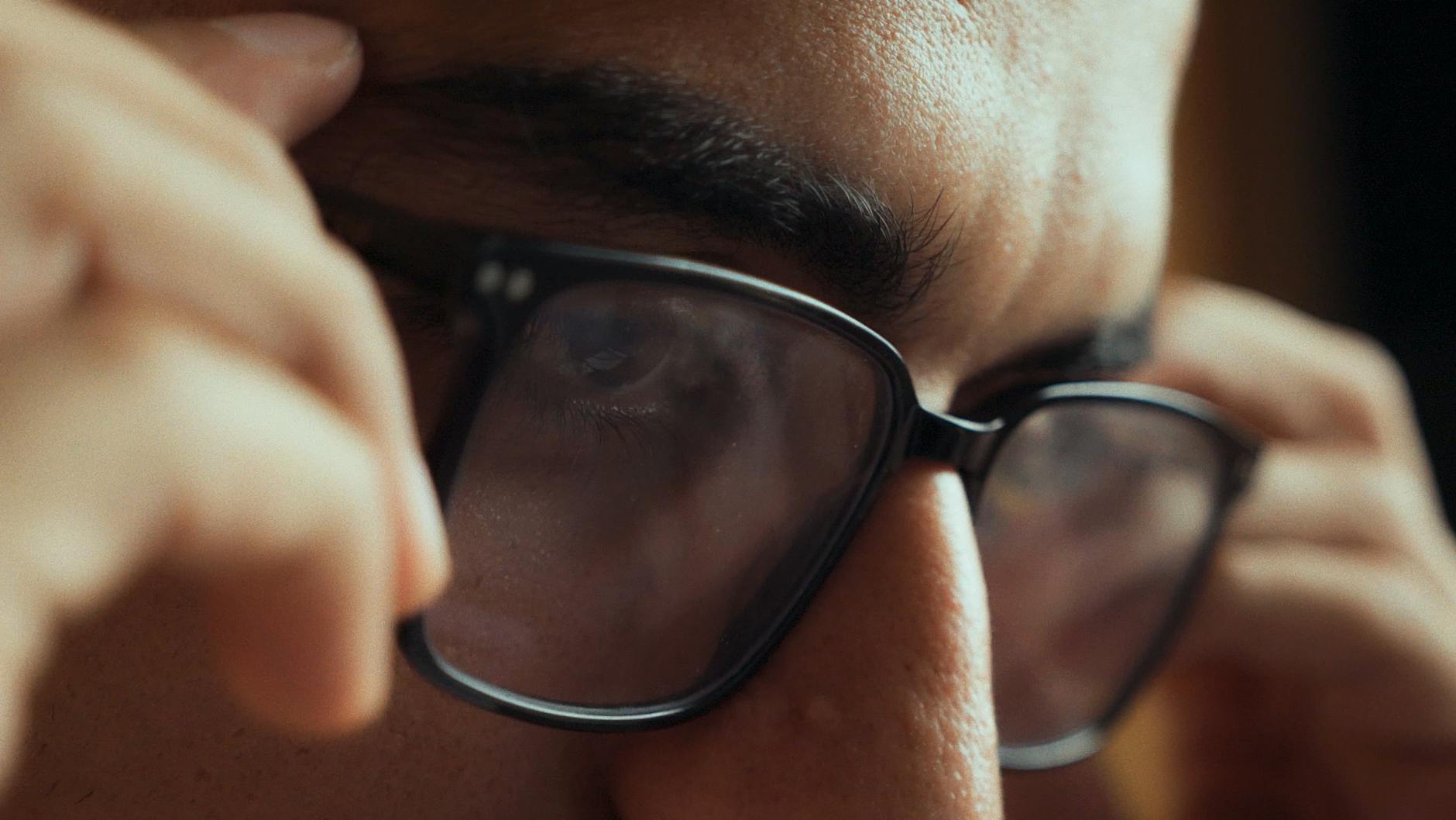
Rubbing Eyes

The mechanical pressure applied during eye rubbing can be surprisingly forceful and is distributed directly onto the cornea and sclera. In individuals with any underlying corneal vulnerability, repeated rubbing accelerates a condition called keratoconus, where the cornea progressively thins and bulges outward. The hands carry significant bacterial and viral loads that are transferred efficiently to the ocular surface with every touch. Eye rubbing also ruptures tiny capillaries around the eye, contributing to persistent redness and puffiness that worsens over time. Itching is better addressed with preservative-free eye drops or a cold compress rather than direct contact.
UV Neglect

Ultraviolet radiation from sunlight reaches the eyes on both sunny and overcast days, as clouds block only a fraction of UV output. Cumulative UV exposure is one of the most well-documented accelerants of cataract formation, causing the eye’s natural lens to yellow and cloud earlier than it otherwise would. Pterygium, a fleshy growth that spreads across the cornea from the white of the eye, is also strongly associated with chronic unprotected UV exposure. Reflected UV from water, sand, and snow intensifies exposure significantly beyond what direct sunlight alone delivers. Wraparound sunglasses rated for full UV400 protection are the most effective daily shield against this damage.
Poor Lighting

Reading or working in lighting that is either too dim or creates harsh glare forces the eye’s pupil and focusing muscles into constant compensatory adjustment. Dim lighting causes sustained pupil dilation, which reduces optical clarity and demands greater muscular effort to maintain a sharp focal point on text or detail. Overhead fluorescent lighting and unshielded bare bulbs create high-contrast glare that generates eye fatigue within minutes of exposure. The ideal working environment uses diffused, warm-toned light sources positioned to illuminate the task without reflecting back into the eyes. Consistently poor lighting environments have been linked to chronic headaches and accelerated accommodative fatigue in working-age adults.
Unprotected Swimming

Chlorinated pool water contains chemical compounds that strip the natural tear film from the corneal surface within seconds of contact. These same compounds disrupt the delicate microbial balance of the ocular surface, weakening its natural defenses against opportunistic pathogens. Open water swimming introduces organic contaminants including bacteria, algae toxins, and amoeba species that have caused severe and permanent vision loss in documented cases. Acanthamoeba keratitis, a rare but devastating corneal infection, is almost exclusively associated with water exposure in contact lens wearers. Sealed swim goggles are the only reliable barrier against these chemical and biological hazards.
Smoking

Tobacco smoke contains over four thousand chemical compounds, many of which enter the bloodstream and directly affect the microvasculature that supplies blood to the retina and optic nerve. Smokers are statistically two to four times more likely to develop age-related macular degeneration compared to non-smokers, according to large-scale population studies. The oxidative stress triggered by tobacco metabolites depletes antioxidant reserves in ocular tissue faster than the body can replenish them. Cataracts also develop earlier and progress more rapidly in consistent smokers than in otherwise comparable non-smoking populations. Even secondhand smoke exposure over prolonged periods measurably increases inflammatory markers within the ocular surface.
Contact Lens Overuse
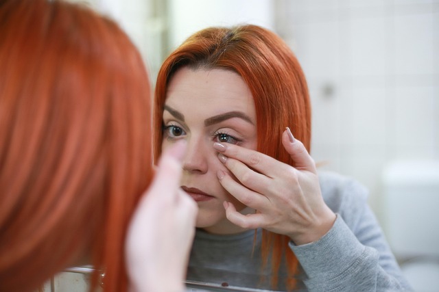
Wearing contact lenses beyond the recommended daily duration restricts oxygen flow to the cornea, which is an avascular tissue that depends on direct atmospheric oxygen. Hypoxic stress from extended contact lens wear stimulates the growth of abnormal blood vessels into the cornea in a process called neovascularization. Sleeping in lenses dramatically multiplies infection risk by creating a warm, moist environment where bacteria multiply rapidly against the corneal surface. Tap water contact with lenses introduces microorganisms that standard lens solutions cannot fully neutralize. Ophthalmologists consistently identify contact lens non-compliance as one of the most preventable causes of serious corneal damage in clinical practice.
Dehydration

The tear-producing glands require adequate systemic hydration to maintain consistent lacrimal output throughout the day. Even mild dehydration reduces tear volume and alters the biochemical composition of the tear film, compromising its protective and lubricating properties. A destabilized tear film exposes the corneal epithelium to environmental irritants and increases friction with every blink. Chronic low-grade dehydration is a frequently overlooked contributing factor in patients presenting with persistent dry eye symptoms that do not fully respond to topical treatments. Adults who consume sufficient water throughout the day demonstrate measurably better tear film stability scores in clinical assessments.
High Sugar Diet

Elevated blood glucose levels trigger a process called glycation, where sugar molecules attach to proteins within the lens and retinal tissues, altering their structural integrity. The lens is particularly vulnerable to glycation-related clouding, which is why diabetic populations develop cataracts at significantly younger ages than the general population. High-sugar diets that produce frequent blood glucose spikes also promote chronic low-level inflammation throughout the body, including within the delicate vascular networks of the retina. Diabetic retinopathy, a leading cause of preventable blindness worldwide, begins with glucose-driven damage to the retinal capillaries long before any visual symptoms appear. Reducing refined sugar intake is therefore one of the most impactful dietary decisions for long-term retinal health.
Vitamin A Deficiency

Vitamin A is an essential structural component of rhodopsin, the photosensitive protein that enables the rod cells in the retina to detect light under low-light conditions. Insufficient dietary intake of this nutrient is the leading preventable cause of blindness in children across low-income regions globally. Even in developed countries, poor dietary patterns that exclude liver, dairy, eggs, and orange and yellow plant foods can produce subclinical deficiency over time. Early symptoms include difficulty adjusting to dim environments, a condition clinically described as nyctalopia or night blindness. Prolonged deficiency progresses to xerophthalmia, a drying and thickening of the conjunctiva that can lead to corneal scarring if left unaddressed.
Omega-3 Deficiency

The meibomian glands lining the eyelids secrete an oily layer that forms the outermost component of the tear film and slows its evaporation between blinks. These glands function most effectively when the body has adequate circulating omega-3 fatty acids, which influence the fluidity and quality of their secretions. Diets low in oily fish, flaxseed, and walnuts are associated with meibomian gland dysfunction, a condition that is now recognized as the most common underlying cause of evaporative dry eye disease. Randomized controlled trials have demonstrated that omega-3 supplementation produces measurable improvements in tear film stability and dry eye symptom scores. The anti-inflammatory properties of these fatty acids also provide broader protective effects within the retinal tissue.
Sleep Deprivation

The eyes undergo critical restorative processes during sleep, including replenishment of tear film components, clearance of metabolic waste from ocular tissues, and repair of cellular damage accumulated during waking hours. Consistently sleeping fewer than seven hours per night is associated with chronic dry eye, persistent redness, and increased light sensitivity that does not fully resolve between days. Intraocular pressure follows a circadian rhythm that sleep disruption destabilizes, which is a relevant concern for individuals with glaucoma or elevated pressure risk. Floppy eyelid syndrome, a condition where eyelid tissue loses structural tone, has a documented association with poor and fragmented sleep patterns. The immune surveillance that protects the ocular surface from infection is also meaningfully suppressed by chronic sleep insufficiency.
Eye Drops Overuse

Over-the-counter redness-relief eye drops work by constricting the blood vessels of the conjunctiva to produce a temporary whitening effect. Regular use of these vasoconstrictive drops creates a rebound dilation effect when the medication wears off, causing eyes to appear redder than before the drop was applied and driving further use. This cycle of dependency can persist for months and may cause permanent changes to the conjunctival vasculature with long-term use. Many popular formulations also contain preservatives that are toxic to the corneal epithelium when applied multiple times daily over extended periods. Preservative-free lubricating drops are the clinically recommended alternative for managing routine dryness and irritation safely.
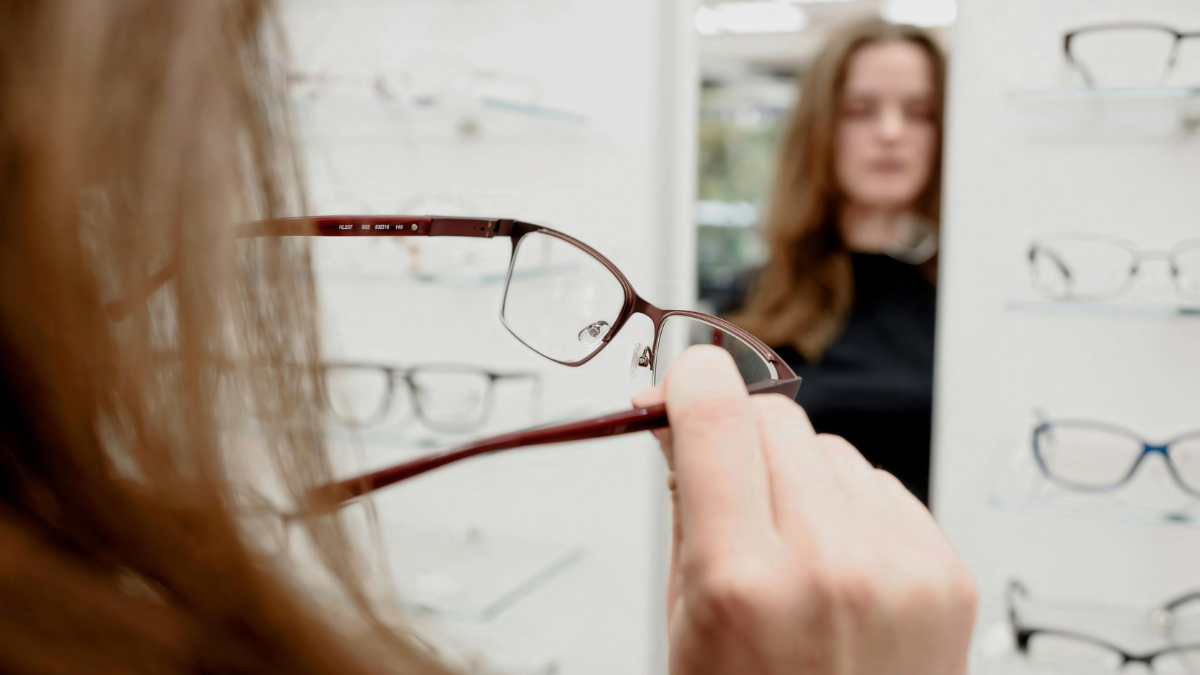
Incorrect Eyewear
Wearing an outdated prescription forces the eyes to compensate for the refractive mismatch through sustained ciliary muscle effort that generates significant fatigue over the course of a day. This visual strain manifests as headaches, difficulty concentrating, and sensitivity to light in many patients who delay updating their corrective lenses. In children, uncorrected or incorrectly corrected refractive errors during critical developmental periods can lead to amblyopia, a form of vision impairment where the brain suppresses input from one eye. Purchasing non-prescription magnification glasses from retail outlets without professional assessment is an equally problematic practice that compounds uncorrected astigmatism and binocular imbalances. Annual professional eye examinations provide the most reliable means of ensuring that corrective prescriptions remain accurate.
High Altitude

At elevations above three thousand meters, ultraviolet radiation intensity increases by approximately ten percent for every thousand meters gained, due to the reduced atmospheric filtering of UV wavelengths. Mountaineers and high-altitude hikers who do not wear appropriate eyewear risk photokeratitis, a painful sunburn of the corneal surface that causes temporary but severe vision impairment. Snow and ice surfaces at altitude can reflect up to eighty percent of incoming UV radiation upward into unprotected eyes, compounding the already elevated exposure. Repeated episodes of photokeratitis cause cumulative corneal scarring that gradually reduces optical clarity. Glacier glasses and wraparound goggles with high UV attenuation ratings are the standard protective equipment for extended time at elevation.
Air Conditioning

Modern air conditioning systems reduce indoor humidity levels significantly, often dropping relative humidity below thirty percent in heavily cooled environments. The corneal tear film evaporates at a measurably faster rate in low-humidity air, producing dry eye symptoms within hours of exposure in susceptible individuals. Air conditioning vents directed at face level create localized airflow that accelerates tear evaporation asymmetrically, often affecting one eye more severely than the other. Office workers who spend eight or more hours per day in heavily air-conditioned environments report some of the highest rates of dry eye disease in occupational health surveys. Humidifiers, strategic vent positioning, and regular lubricating drops provide practical mitigation for this pervasive environmental stressor.
Tight Neckwear

Research published in ophthalmology literature has demonstrated that tightly fastened neckwear such as fitted collars and neckties raises intraocular pressure by restricting venous drainage from the head and neck. This pressure elevation occurs within minutes of applying constricting neckwear and can persist for the entire duration it is worn. For the general population this effect is transient and clinically insignificant, but for individuals with glaucoma or elevated baseline intraocular pressure it represents a meaningful daily risk factor. The jugular vein compression mechanism involved is well understood and correlates directly with collar tightness rather than tie style or material. Optometrists advise patients undergoing tonometry testing to loosen collar buttons beforehand to obtain accurate baseline pressure readings.
Late Eating

Consuming large meals within two to three hours of sleep onset disrupts the metabolic and hormonal environment during the overnight period when ocular repair processes are most active. Elevated postprandial blood glucose from late-night eating produces a prolonged glycemic response that generates oxidative stress in retinal microvasculature during the critical nighttime restoration window. Acid reflux triggered by recumbent digestion has been associated in clinical literature with a condition called nonarteritic anterior ischemic optic neuropathy, where blood supply to the optic nerve is transiently interrupted. Insulin fluctuations during nighttime hours also interfere with the fluid dynamics of the eye, contributing to variable intraocular pressure readings in individuals who eat heavily before bed. Aligning meal timing with earlier evening hours supports the broader metabolic stability that long-term ocular health depends upon.
If any of these habits resonated with your daily routine, share which ones surprised you most in the comments.